|
Case Report
Endoscopic management of fish bone perforation of the 3rd portion of the duodenum leading to retroperitoneal abscess: A case report and literature review
1 Surgical Senior Resident Medical Officer, Department of Surgery, Westmead Hospital, Sydney, NSW, Australia
2 Surgical Registrar, Department of Surgery, Tamworth Base Hospital, Tamworth, NSW, Australia
3 Consultant General Surgeon, Department of Surgery, Tamworth Base Hospital, Tamworth, NSW, Australia
Address correspondence to:
Kah Ann Ho
Westmead Hospital, Westmead, NSW,
Australia
Message to Corresponding Author
Article ID: 100140Z12KH2024
Access full text article on other devices

Access PDF of article on other devices

How to cite this article
Ho KA, Chen K, Srinivasan R. Endoscopic management of fish bone perforation of the 3rd portion of the duodenum leading to retroperitoneal abscess: A case report and literature review. J Case Rep Images Surg 2024;10(2):5–9.ABSTRACT
Introduction: Most fish bones ingested pass through the gastrointestinal tract without complications. Perforation occurs in less than 1% of cases but this rate increases up to 35% with ingestion of sharp pointed objects. There are only 14 cases of duodenal perforation from fish bone described in the literature thus far. In most of these cases there was no history of ingestion reported which makes diagnosis challenging. Complications from such perforation include hepatic abscess, pancreatic abscess, renal vein thrombus, duodenocaval fistula, and lodgement in common bile duct. 25% of these cases were managed with endoscopy alone without need for surgery.
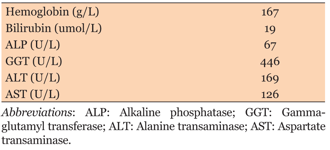
Case Report: We describe a case of a 70-year-old male with perforation involving the third part of the duodenum from a fish bone complicated by retroperitoneal collection. He presented with right-sided abdominal pain and raised inflammatory markers. Initial imaging revealed a foreign body lodged in the third portion of the duodenum with adjacent retroperitoneal fluid. Progress imaging showed new extraluminal gas and inflammation prompting urgent intervention. Endoscopic removal of the fish bone was successful with subsequent resolution of the abscess.
Conclusion: In this case, endoscopic removal of the fish bone was successful resulting in early recovery. However, management of duodenal perforation by foreign body can be challenging due to its rarity and myriad of possible management options including conservative, percutaneous, endoscopic, and surgical approaches.
Keywords: Duodenal perforation, Endoscopy, Fish bone perforation, Foreign body, Retroperitoneal abscess
SUPPORTING INFORMATION
Author Contributions
Kah Ann Ho - Substantial contributions to conception and design, Analysis of data, Drafting the article, Revising it critically for important intellectual content, Final approval of the version to be published
Kabytto Chen - Acquisition of data, Drafting the article, Final approval of the version to be published
Rajkumar Srinivasan - Substantial contributions to conception and design, Acquisition of data, Analysis of data, Interpretation of data, Drafting the article, Revising it critically for important intellectual content, Final approval of the version to be published
Guarantor of SubmissionThe corresponding author is the guarantor of submission.
Source of SupportNone
Consent StatementWritten informed consent was obtained from the patient for publication of this article.
Data AvailabilityAll relevant data are within the paper and its Supporting Information files.
Conflict of InterestAuthors declare no conflict of interest.
Copyright© 2024 Kah Ann Ho et al. This article is distributed under the terms of Creative Commons Attribution License which permits unrestricted use, distribution and reproduction in any medium provided the original author(s) and original publisher are properly credited. Please see the copyright policy on the journal website for more information.