|
Case Report
A rare presentation of colonic malignancy: Enterocutaneous fistula secondary to locally advanced cancer
1 Unaccredited Surgical Registrar, Department of Surgery, Gladstone Hospital, Gladstone, QLD, Australia
2 Director of Surgery, Department of Surgery, Gladstone Hospital, Gladstone, QLD, Australia
3 Pathology Registrar, Institute of Clinical Pathology and Medical Research, Westmead, NSW, Australia
Address correspondence to:
Neesa Fadaee
58 Wiare Circuit Orange, NSW 2800,
Australia
Message to Corresponding Author
Article ID: 100059Z12NF2019
Access full text article on other devices

Access PDF of article on other devices


How to cite this article
Fadaee N, De Clercq S, Fadaee S. A rare presentation of colonic malignancy: Enterocutaneous fistula secondary to locally advanced cancer. J Case Rep Images Surg 2019;5:100059Z12NF2019.ABSTRACT
Introduction: Colorectal cancer is the third most common cancer in the world. It is usually identified during screening programs, however some patients present to emergency departments with symptoms such as hemorrhage, perforation, or bowel obstruction. Locally advanced presentations of colorectal cancer are rare.
Case Report: A 69-year-old male presented to our hospital with a large left lumbar wound with active feculent discharge and associated cellulitis. He did not report any other symptoms. Radiological investigations suggested an enterocutaneous fistula with an associated anterior abdominal wall collection. A flexible sigmoidoscopy was performed revealing a fungating tumor in the sigmoid colon. Unfortunately, the patient developed significant per rectal bleeding and blood transfusion dependency. He was palliated on the ward following a multidisciplinary family meeting.
Conclusion: Enterocutaneous fistula formation secondary to locally advanced colorectal cancer is quite rare. There have only been five previously published case reports. The management of such cases is quite complex as it requires a multidisciplinary approach and there are no treatment or prognosis guidelines currently. Our case is of particular interest as it is the first reported case in a developed country.
Keywords: Colorectal cancer, Emergency presentation, Enterocutaneous fistula
Introduction
Colorectal cancer can present in many ways. Most cancers are identified with screening programs [1]. Despite this about a third of colorectal cancer presents to the emergency department with symptoms requiring urgent intervention [1],[2]. These commonly include hemorrhage, bowel obstruction, or perforation [2]. The development of an enterocutaneous fistula typically occurs in the context of an inflammatory condition such as inflammatory bowel disease or diverticulitis, and is only ever rarely associated with colorectal cancer [1],[2]. Indeed, there have only been five previously published case reports describing this presentation [3],[4],[5],[6],[7]. As can be expected, the management of these cases is difficult as the formation of an enterocutaneous fistula represents locally advanced disease [3]. There are currently no guidelines dictating treatment modalities and limited data regarding prognosis [4].
Case Report
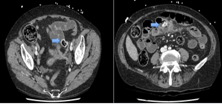
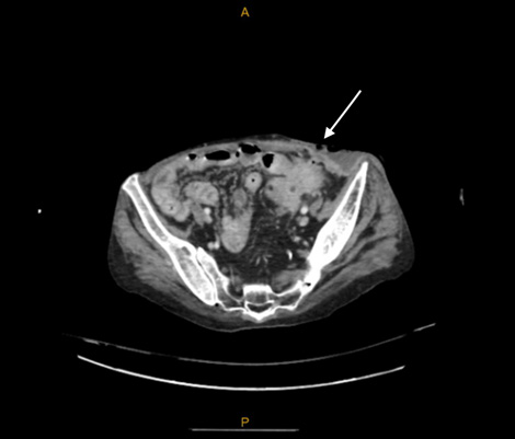
A 69-year-old male presented to our hospital after his neighbors alerted ambulance services due to concerns regarding his welfare. On presentation he was noted to have a large, left lumbar abdominal wound, measuring approximately 10 cm (Figure 1). The wound appeared to have a necrotic center with associated surrounding cellulitis and was discharging feculent material.Despite the presence of this wound, he appeared otherwise systemically well. In fact, no constitutional symptoms were elicited through a comprehensive system review. The patient denied any significant medical comorbidities and did not take any regular medications. He did, however, report being an active smoker and a history of excessive alcohol intake. Perusal of his past history gleaned that he underwent a colonoscopy five years ago, with identification and subsequent piecemeal resection of a sigmoid polyp. Histopathological examination of this polyp revealed a tubular adenoma with high grade dysplasia. Repeat endoscopy in 12 months was planned, however the patient was unfortunately lost to follow-up. Physical examination of the patient revealed an elderly man with all vital signs, including temperature, within normal limits. He appeared emaciated with a large, left lumbar wound. Despite the wound, the patient’s abdomen was soft and non-tender with no evidence of localized or generalized peritonitis. The rest of the examination was essentially normal, with no other significant positive findings of note. The patient then proceeded to a computed tomography (CT) scan of his abdomen and pelvis which revealed a short segment of abnormal mucosal thickening and enhancement of the sigmoid colon with a thin, linear fistula connecting to the superficial abdominal wall (Figure 2). The appearance was consistent with an enterocutaneous fistula with an anterior abdominal collection. Additionally, a focal polypoid enhancing lesion was noted in the anterior wall of the rectum. There was no radiological evidence of diverticular disease.The patient was noted to have raised white cell count of 19.8×10⁹/L (4.0–11.0×10⁹/L) with an associated neutrophilia. His hemoglobin was 178 g/L (135–180 g/L) and renal function was within normal limits. Liver function was also preserved; however, the patient’s albumin was noted to be 25 g/L (35–50 g/L), fitting with his emaciated appearance. A colostomy bag was placed over the fistula and the patient was commenced on intravenous piperacillin and tazobactam. A flexible sigmoidoscopy was subsequently performed to further investigate the rectal lesion and discovered two polypoid lesions in addition to a fungating tumor, which was biopsied. Histopathological examination of the biopsy specimen demonstrated a tubular adenoma with high grade dysplasia with findings suspicious of an invasive adenocarcinoma. The patient was subsequently found to have a positive blood culture with growth of Proteus mirabilis in one of two bottles after 31.5 hours. Antibiotic sensitivity testing demonstrated appropriate sensitivity to piperacillin and tazobactam. On day 3 of admission, the patient developed intractable hematochezia with an associated significant drop in his hemoglobin necessitating considerable transfusion support and, ultimately, transfusion dependency. Due to the severity of the patient’s clinical condition, consultation between the treating medical teams and patient’s family leads to a consensus opinion that palliation was the most appropriate management strategy. The patient subsequently passed away a few days following this.

Discussion
Colorectal cancer is the third most common cancer in the world, and the fourth most common cause of cancer death [1]. Patients with bowel cancers typically present for routine surgical review following the development of often non-specific symptoms such as changes in bowel habits, abdominal pain, and bloating of PR bleeding [2]. Emergency department presentations, on the other hand, tend to include large bowel obstruction, perforation, and bleeding [2]. However, locally advanced presentations of colorectal cancer are rare. Indeed, the presenting feature in this case is an unusual one with only five previously published case reports of patients with a colorectal cancer presenting with an enterocutaneous fistula [3],[4],[5],[6],[7]. In all previous cases, the patients had a similarly delayed presentation to a health service; however, these cases all occurred in developing countries. Table 1 summarizes the five previously published case reports (Table 1). Of these five cases, four patients developed enterocutaneous fistulae involving their abdominal walls and one patient developed a fistula on their back. All the cases occurred in the context of a primary colonic malignancy and surgical resection with curative intent was performed [7]. The development of an enterocutaneous fistula in patients with colorectal cancer indicates locally advanced disease. The pathophysiology underpinning fistula formation has been postulated to be due to localized perforation with delayed presentation leading to locoregional extension and, thus, cutaneous invasion of the colorectal cancer [6]. There are currently no guidelines regarding the management and prognosis of such complications, presumably due to the relative paucity of cases [4],[6]. Nevertheless, management of these cases is complicated and requires a multidisciplinary approach. As mentioned, in the previously published case studies, all patients underwent a bowel resection with the aim of definitive surgical management and subsequent closure of the enterocutaneous fistula; surgical management should certainly be considered in cases where the patient is stable, can withstand surgical resection with curative intent, and is likely to benefit from the procedure [2]. However, as with any oncological surgery, the patient’s comorbidities and potential improvements to quality of life must be considered.Unfortunately, patients presenting with such advanced disease typically have a higher morbidity and mortality, which complicates or inhibits emergency surgical management [4]. Our case differed from the other published cases as it was complicated by the rapid development of intractable PR bleeding and the subsequent development of transfusion dependency, which precluded surgical management. In the case presented by Gunay et al. the patient also presented with PR bleeding, however this was on a background of known hemorrhoids and, thus, did not preclude operative management [5]. There have been numerous case reports published about unusual initial presentations of locally advanced colorectal cancer, including abdominal wall cellulitis [8], abdominal wall abscess [9], cutaneous invasion [10], and spontaneous enterocutaneous fistula. Spontaneous enterocutaneous fistula formation is commonly associated with diverticular disease, inflammatory bowel disease, and anastomotic breakdown [11]. Colonic malignancy is an uncommon cause of spontaneous enterocutaneous fistula(s) and should be considered as a rare differential diagnosis.

Conclusion
We present our case of spontaneous enterocutaneous fistula secondary to locally advanced colorectal cancer. Our case is of particular interest as it appears to be the first documented case in a developed country and the first published case in which complications impeded surgical management. Colonic malignancy is a rare cause of spontaneous enterocutaneous fistula and further research is required to guide investigation and timely management.
REFERENCES
1.
Arnold M, Sierra MS, Laversanne M, Soerjomataram I, Jemal A, Bray F. Global patterns and trends in colorectal cancer incidence and mortality. Gut 2017;66(4):683–91. [CrossRef]
[Pubmed]

2.
Baer C, Menon R, Bastawrous S, Bastawrous A. Emergency presentations of colorectal cancer. Surg Clin North Am 2017;97(3):529–45. [CrossRef]
[Pubmed]
3.
Nagaraja R, Kudva A, Prasad NN. Spontaneous colocutaneous fistula: A rare presentation of colon carcinoma. Internet J Surg 2008;15:1.
4.
Jethwani U, Bansal AA, Kandwal VV. Spontaneous enterocutaneous fistula due to colonic malignancy: A rare case report. Arch Int Surg 2013;3(2):147–9. [CrossRef]
5.
Günay Y, Bırcan H, Demıralay E, Sade Ç, Emek E, Demırağ A. Spontaneous enterocutaneous fistula: Unusual presentation of colon cancer. Turk J Gastroenterol 2013;24(5):458–9. [CrossRef]
[Pubmed]
6.
Wadhwani N, Diwakar DK. Localised perforation of locally advanced transverse colon cancer with spontaneous colocutaneous fistula formation: A clinical challenge. BMJ Case Rep 2018;2018. pii: bcr-2018-224668. [CrossRef]
[Pubmed]
7.
Kumar A, Pahwa H, Srivastava R, Kumar R. Multiple spontaneous enterocutaneous fistulae on back: A rare presentation of colonic malignancy. BMJ Case Rep 2012;2012. pii: bcr 2012007119. [CrossRef]
[Pubmed]
8.
Bischoff K, López C, Shaffer K, Schwaitzberg S. Colorectal adenocarcinoma presenting as abdominal wall cellulitis. Radiol Case Rep 2015;3(3):204. [CrossRef]
[Pubmed]
9.
Kargar S, Arjmandzadeh E, Ehsani F, sadat Alavizadeh F, Talebi S. Atypical presentation of sigmoid colon carcinoma as right lower quad-rant abdominal wall abscess: the visible part of iceberg. Journal of Case Reports in Practice (JCRP) 2017;5(1):4–6.
10.
Tenreiro N, Ferreira C, Silva S, et al. Locally advanced colon cancer with cutaneous invasion: Case report. BMC Res Notes 2017;10(1):113. [CrossRef]
[Pubmed]
11.
Schecter WP. Management of enterocutaneous fistulas. Surg Clin North Am 2011;91(3):481–91. [CrossRef]
[Pubmed]
SUPPORTING INFORMATION
Author Contributions
Neesa Fadaee - Conception of the work, Design of the work, Acquisition of data, Analysis of data, Drafting the work, Revising the work critically for important intellectual content, Final approval of the version to be published, Agree to be accountable for all aspects of the work in ensuring that questions related to the accuracy or integrity of any part of the work are appropriately investigated and resolved.
Stefaan De Clercq - Conception of the work, Design of the work, Revising the work critically for important intellectual content, Final approval of the version to be published, Agree to be accountable for all aspects of the work in ensuring that questions related to the accuracy or integrity of any part of the work are appropriately investigated and resolved.
Shannon Fadaee - Analysis of data, Drafting the work, Revising the work critically for important intellectual content, Final approval of the version to be published, Agree to be accountable for all aspects of the work in ensuring that questions related to the accuracy or integrity of any part of the work are appropriately investigated and resolved.
Guarantor of SubmissionThe corresponding author is the guarantor of submission.
Source of SupportNone
Consent StatementWritten informed consent was obtained from the patient for publication of this article.
Data AvailabilityAll relevant data are within the paper and its Supporting Information files.
Conflict of InterestAuthors declare no conflict of interest.
Copyright© 2019 Neesa Fadaee et al. This article is distributed under the terms of Creative Commons Attribution License which permits unrestricted use, distribution and reproduction in any medium provided the original author(s) and original publisher are properly credited. Please see the copyright policy on the journal website for more information.