|
Case Report
Foramen of Winslow hernia in a patient with ventriculoperitoneal shunt: A case report and review of literature
1 FRACS, Locum Staff Specialist in Surgery, Department of Surgery, Caboolture Hospital, Metro North Health Services, 87/129 McKean Street, Caboolture, Queensland, Australia
2 MRCS (Edinburgh), Senior surgical trainee, Department of Surgery, Caboolture Hospital, Metro North Health Services, 87/129 McKean Street, Caboolture, Queensland, Australia
3 MD (Iran), Principal House Officer, Department of Surgery, Caboolture Hospital, Metro North Health Services, 87/129 McKean Street, Caboolture, Queensland, Australia
4 MD (Damascus University), Principal House Officer, Department of Surgery, Caboolture Hospital, Metro North Health Services, 87/129 McKean Street, Caboolture, Queensland, Australia
5 MBBS, Intern, Department of Surgery, Caboolture Hospital, Metro North Health Services, 87/129 McKean Street, Caboolture, Queensland, Australia
6 FRACS, Staff Specialist in Surgery, Acting Director of Surgery, Department of Surgery, Caboolture Hospital, Metro North Health Services, 87/129 McKean Street, Caboolture, Queensland, Australia
Address correspondence to:
Prasenjit Raychaudhuri
6 Harley Place, Kellyville, Sydney, New South Wales, Post Code 2155,
Australia
Message to Corresponding Author
Article ID: 100173Z12PR2026
Access full text article on other devices

Access PDF of article on other devices


How to cite this article
Raychaudhuri P, Seelamanthula VVR, Motamedi MJ, Fadel A, Elege AA, Hendahewa R. Foramen of Winslow hernia in a patient with ventriculoperitoneal shunt: A case report and review of literature. J Case Rep Images Surg 2026;12(2):14–20.ABSTRACT
Introduction: Foramen of Winslow hernias are a rare subset of internal hernias. They are difficult to diagnose clinically. They should be considered by surgeons in the differential diagnosis in patients presenting with abdominal pain. A high index of suspicion and early imaging are necessary for prompt diagnosis and treatment to avoid morbidity and mortality.
Case Report: A 73-year-old patient presented to the emergency department with complaints of severe epigastric pain that had significantly reduced by the time of presentation. She had a ventriculoperitoneal shunt for hydrocephalus about four years ago. An urgent multidetector computed tomography scan performed in the emergency department demonstrated an internal hernia, with the cecum herniating through the foramen of Winslow. Emergency surgery was commenced laparoscopically but was converted to laparotomy as the herniated contents could not be reduced laparoscopically. The patient made an uneventful postoperative recovery and was discharged home on the fifth postoperative day.
Conclusion: Urgent surgical intervention is necessary in these cases. There is a lack of clear guidelines for abdominal surgery in the presence of a ventriculoperitoneal shunt. A neurosurgical opinion should be sought, preferably before proceeding to surgery. There is debate regarding the specifics of surgical management of these hernias, with a lack of clear guidelines.
Keywords: Emergency surgery, Foramen of Winslow, Internal hernia, Neurosurgery
Introduction
Internal hernias (IHs) are a rare cause of intestinal obstruction, accounting for 0.6% to 5.5% of cases [1],[2]. To the best of our knowledge, this is the first reported case of foramen of Winslow hernia (FWH) in the presence of a ventriculoperitoneal shunt (VPS).
Foramen of Winslow hernias constitute 8% of IHs [3],[4]. Internal hernias can occur through congenital or acquired openings in the peritoneal cavity. They can occur through normal peritoneal orifices or congenital defects resulting from developmental anomalies, or through acquired internal openings secondary to inflammation, trauma, or surgery. The foramen of Winslow (FW) is a normal opening in the peritoneal cavity. Foramen of Winslow hernia can therefore be subcategorized as a normal-aperture subtype of congenital IH [5],[6],[7].
In the past, IHs were rarely detected preoperatively and were often detected during laparotomy [8]. As they are difficult to diagnose clinically, imaging plays a significant role in early diagnosis. Multidetector computed tomography (MDCT) has become an important tool for the early detection of these rare hernias [6],[9]. With increasing preoperative detection, surgery can be performed before the onset of ischemia of the herniated intestines.
Due to a lack of defined guidelines, there is debate about the most appropriate surgical management of these rare hernias [10]. While traditionally managed by laparotomy, increasingly, laparoscopic and, in a few cases, robotic approaches have been used [10],[11].
Case Report
A 73-year-old female presented to the emergency department with severe overnight epigastric pain radiating to the shoulders. On presentation to the emergency department, her pain had reduced and clinical findings were non-specific.
Her past medical history included a subarachnoid hemorrhage due to rupture of a cerebral aneurysm, with hydrocephalus necessitating the insertion of a VPS about four years ago at another hospital. The only abdominal surgery was a small transverse incision on the right abdomen at the level of the umbilicus for insertion of the VPS. She had also undergone pelvic bone fracture repair following a motor vehicle accident many years ago and a total hip replacement in the past.
On examination, her blood pressure was 180/84 with other vital signs being normal. Her hemoglobin, white cell counts, electrolytes, liver, and renal function tests were all normal. Physical examination demonstrated mild epigastric distension with mild tenderness in the epigastric region. There was no evidence of peritonitis.
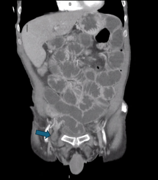
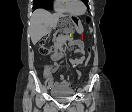
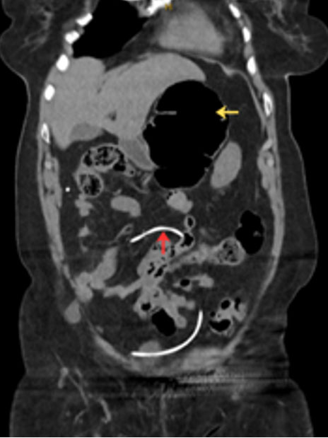
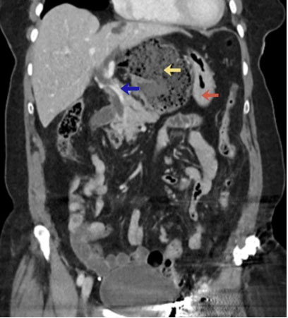
A contrast-enhanced MDCT with angiogram performed in the emergency department showed caecum passing through the FW and lying in the lesser sac (Figure 1). The cecum was obstructed and dilated with no signs of pneumatosis intestinalis. The distal catheter of the VPS was noted in the peritoneal cavity (Figure 2). Partial effacement of the main portal vein (PV) was noted (Figure 3). The coeliac trunk, superior mesenteric artery and inferior mesenteric artery were patent with no significant stenosis.
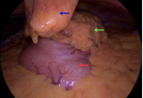
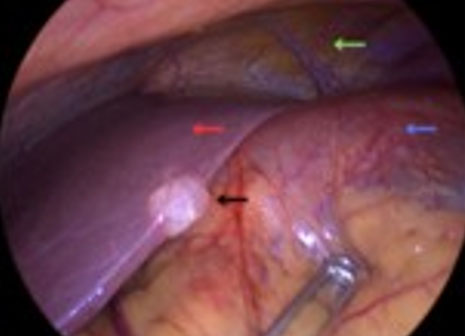
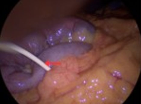
She was taken to the operating theatre for emergency surgery. A laparoscopic approach was attempted first. A 10 mm subumbilical port, and 5 mm right and left upper abdomen ports were inserted. The cecum was found to be herniating through the FW into the lesser sac (Figure 4 and Figure 5). The distal catheter of the VPS was also noted in the peritoneal cavity (Figure 6). Attempts were made to reduce the contents of the hernia with gentle traction using laparoscopic graspers. However, reduction was difficult and the decision was made to convert the procedure to laparotomy. An upper midline laparotomy was performed and, after gentle bimanual manipulation, the herniated cecum and small intestine were reduced. The cecum and proximal ascending colon were edematous and redundant but viable. The FW allowed entry of the index and middle fingers suggesting a diameter of approximately 3 cm. At this stage, a cecopexy was considered, but it was decided not to perform fixation (pexy). The distal catheter of the VPS was carefully handled throughout the procedure. The patient had an uneventful postoperative recovery and at 3-week postoperative follow-up, she was progressing well, with an uncomplicated recovery.
Discussion
A clear understanding of anatomy is necessary for various possible internal hernias. The FW is the communication between the greater and lesser sac in the peritoneal cavity. Its anterior border is the free edge of the lesser omentum (containing the portal triad), the posterior border is formed by the inferior vena cava. The superior and inferior borders are formed by the caudate process of the liver and the duodenum, respectively [2]. Predisposing factors for IH through this opening are unclear. Those mentioned in the literature include increased visceral mobility, enlargement of the foramen of Winslow (>3 cm) and an increase in intra-abdominal pressure [2],[10],[12]. Further research is needed to elucidate the role of these predisposing factors [13].
To create a nuanced approach to radiological diagnosis of these rare IH, attempts have been made to categorize them into groups. Doishita [9] mentions classification of internal hernias into three main types, based on herniation into a normal foramen, an unusual peritoneal fossa or recess, or through an abnormal opening in the mesentery or peritoneal ligament. Myers [4], using historic data, broadly classified IH as paraduodenal (53%), pericecal (13%), FW (8%), transmesenteric, transmesocolic (8%), intersigmoid (6%), and retroanastomotic (5%). The remaining 7% include paravesical hernias, which are not considered to represent true internal hernias by other authors.
Differential diagnosis of abdominal pain is often challenging for surgeons. Foramen of Winslow is a rare subset of IH [7],[12],[14]. Accurate preoperative diagnosis of internal hernias is difficult to achieve clinically, and in many cases, diagnosis has been made at laparotomy, laparoscopy, or even autopsy [8],[15],[16],[17]. Thorough knowledge of IH is necessary for surgeons, and they should be considered in the differential diagnosis of abdominal pain. Vague physical signs with subtle and non-specific clinical findings can be misleading and makes it difficult to make an accurate preoperative diagnosis [1],[15],[16],[18],[19]. Foramen of Winslow hernia detected on preoperative MDCT has been noted to have reduced spontaneously during surgical intervention [11],[14]. These hernias can be persistent or cause only intermittent symptoms due to spontaneous reduction. They may also be completely asymptomatic, detected incidentally during surgery for unrelated conditions. This wide range of clinical presentations makes it difficult to arrive at an accurate diagnosis [9],[20].
In various IH, early imaging and diagnosis can prevent ischemia and necrosis of herniated contents [5]. Multidetector computed tomography scan has become an important tool and created a paradigm shift in the early diagnosis and management of these hernias [6],[9]. However, Tatagari [17] reported a case where the diagnosis of FWH was missed even on MDCT, and the patient was taken to theatre for laparoscopy with a provisional diagnosis of cholecystitis. The hernia was detected during laparoscopy.
There is controversy and lack of agreement about the surgical management of these hernias [10],[11],[15],[16],[21],[22],[23]. Rarely, conservative management with spontaneous reduction of FWH has been reported in a patient with multiple comorbidities [21]. However, early detection and prompt surgical intervention are the treatment of choice [1],[2],[22],[23]. Due to the rarity of these hernias and the lack of established guidelines, various surgical techniques have been used. Reduction of hernial contents through the narrow FW has been possible by bimanual reduction in open cases and laparoscopically or robotically in others [10],[11],[23],[24]. In some reports, decompression of the cecum has been necessary before reduction could be achieved [2]. In some reports, a preliminary resection of herniated contents has been necessary prior to reduction during laparotomy [7]. In cases performed laparoscopically, cecal decompression to reduce FWH has been reported in 2 out of 15 cases [10]. In 1 out of 2 cases done performed robotically, cecal decompression was necessary [11].
Prophylactic measures like fixation of the caecum or resection of the redundant colon undertaken to prevent recurrence of FWH are controversial [7],[10],[12],[14],[15],[16],[18],[23]. Clear uniformity of approach is lacking. The wide variety of prophylactic measures that have been used or not used in surgical treatment reflects the differences in opinions about repair techniques. Following reduction, resection has been done for non-viable contents in 2 out of 15 reports of laparoscopic repairs and in some cases during laparotomy [2],[10],[16],[23]. Some authors have performed right hemicolectomy for redundancy of the caecum in the absence of any signs of ischemia or gangrene, to prevent future herniation [17]. Some surgeons have performed fixation (“pexy”) of the viable caecum and right colon [22]. Others have not deemed it necessary to perform resection or any form of fixation of the caecum or right colon where contents appeared viable [12],[13],[14].
The practice of prophylactic closure of FW is not uniform with differing viewpoints among surgeons. Some authors have closed the FW [13],[16],[18],[22]. Many surgeons have chosen not to close the foramen [2],[12],[14],[15],[17]. Out of 15 cases performed laparoscopically, in four cases FW closure was done, one patient underwent FW closure with cecopexy. In nine cases FW was not closed and it was not reported in one [10]. Foramen of Winslow was closed in all 3 cases reported by Cherian [11]. Omental patch has been frequently used to reduce the size of the foramen [11],[12],[18],[22]. Various suturing techniques have also been used to close FW [13],[16]. Table 1 details the various approaches used in the surgical management of these hernias.
Closure of FW can have serious consequences with risks of portal vein thrombosis, bile duct injury and damage to the hepatic artery [14],[15]. Postoperative fibrosis has been postulated to cause reduction of the size of FW. Therefore, the necessity of closure of FW with attendant risks is disputed. This issue has been further confounded by the fact that irrespective of whether prophylactic measures were taken or not, no case of recurrence of these hernias have been reported [2],[7],[11],[14],[15],[22],[23]. Larger studies are necessary to provide clearer evidence regarding the surgical management of these hernias [13]. At this stage, it is difficult to assign clear roles to the various prophylactic surgical techniques that have been described in the literature to prevent recurrence of these rare hernias.
While open repair with laparotomy for IH was common in the past, increasingly laparoscopic and robotic repairs are being reported [8],[10],[11]. In a review of the literature on laparoscopic management of FWH, Morris [10] found 15 cases treated laparoscopically. Cherian [11] performed robotic repair in 2 out of 3 cases of FWH, laparotomy was done in one patient. Others started with laparoscopy and converted to laparotomy as reduction of the hernia was not possible laparoscopically [16],[17],[24].
In our opinion, where possible, laparoscopy should be the first line of treatment in the absence of evidence of ischemia or necrosis of herniated contents. Laparoscopy affords clear views and anatomical assessment with the usual benefits of minimally invasive surgery. Conversion to laparotomy may be necessary to prevent injuries to the hernia contents from forceful reduction. Gentle handling is necessary to prevent inadvertent damage and perforation of contents. We decided to convert to laparotomy as excessive instrumental traction to reduce contents was becoming necessary. In our case, there was significant concern about inadvertent enterotomies and spillage of contents leading to infection of the VPS. As the herniated contents were viable, we did not need to perform any intestinal resections. We decided against fixation techniques due to concerns about potential damage to the cecal or colonic wall with leakage of contents. Besides, there is a lack of any clear evidence of the benefit of fixation of the colon in preventing future recurrences.
The presence of VPS can create challenges during abdominal surgery as most surgeons are unfamiliar with them due to the rarity of such events. However, central nervous system infection following abdominal surgery in the presence of a VPS is rarely reported in the literature. Neurosurgery consultation prior to surgery has been suggested [25]. In our case, prior neurosurgery guidance was obtained. However, as expected, the advice was very general.

Conclusion
Foramen of Winslow hernia is rare and presents with subtle findings. Early diagnosis and treatment have been challenging. Prompt surgical intervention is the preferred approach to management. With the advent of MDCT, laparoscopic and robotic surgery, the approaches to surgical management of these hernias are evolving. However, many aspects of treatment, such as the role of preventive measures, are controversial and need further clarification through larger studies.
REFERENCES
1.
Ge Q, Cong P. CT imaging of a rare combined internal hernia traversing the lesser sac via the gastrocolic ligament and foramen of Winslow. Egypt J Radiol Nucl Med 2025;56:58. [CrossRef]

2.
Sammut M, Barben C. Herniation of the caecum and ascending colon through the foramen of Winslow: A case report and review. Ann R Coll Surg Engl 2020;102(2):e39–41. [CrossRef]
[Pubmed]
3.
Salar O, El-Sharkawy AM, Singh R, Speake W. Internal hernias: A brief review. Hernia 2013;17(3):373–7. [CrossRef]
[Pubmed]
4.
Meyers MA. Dynamic radiology of the abdomen: Normal and pathologic anatomy. 4th ed. New York, NY: Springer-Verlag; 1994.
5.
Lanzetta MM, Masserelli A, Addeo G, Cozzi D, Maggialetti N, Danti G, et al. Internal hernias: A difficult diagnostic challenge. Review of CT signs and clinical findings. Acta Biomed 2019;90(5-S):20–37. [CrossRef]
[Pubmed]
6.
Martin LC, Merkle EM, Thompson WM. Review of internal hernias: Radiographic and clinical findings. AJR Am J Roentgenol 2006;186(3):703–17. [CrossRef]
[Pubmed]
7.
Carpenter SL, Campbell J, Jayaraman V. Caecal volvulus in a foramen of Winslow hernia. BMJ Case Rep 2022;15(3):e247316. [CrossRef]
[Pubmed]
8.
Newsom BD, Kukora JS. Congenital and acquired internal hernias: Unusual causes of small bowel obstruction. Am J Surg 1986;152(3):279–85. [CrossRef]
[Pubmed]
9.
Doishita S, Takeshita T, Uchima Y, Kawasaki M, Shimono T, Yamashita A, et al. Internal hernias in the era of multidetector CT: Correlation of imaging and surgical findings. Radiographics 2016;36(1):88–106. [CrossRef]
[Pubmed]
10.
Moris D, Tsilimigras DI, Yerokun B, Seymour KA, Guerron AD, Fong PA, et al. Foramen of Winslow hernia: A review of the literature highlighting the role of laparoscopy. J Gastrointest Surg 2019;23(10):2093–9. [CrossRef]
[Pubmed]
11.
Cherian HM, Lomas C, Khosravani N, Polcz M, Rabaza JR. Foramen of Winslow hernias: A case series of a rare cause of bowel obstruction in three women. Cureus 2025;17(9):e92729. [CrossRef]
[Pubmed]
12.
Srisajjakul S, Vuttipitayamongkol W, Bangchokdee S. Foramen of Winslow hernia: A rare condition requiring clinical vigilance. J Gastrointest Surg 2025;29(1):101863. [CrossRef]
[Pubmed]
13.
Krishnan A, Mackay TG, Iyengar K, Joel A. Unveiling the foramen of Winslow: A case report of internal hernia and its surgical implications. Cureus 2025;17(8):e90562. [CrossRef]
[Pubmed]
14.
Tani D, Fry D. Caecal herniation through the foramen of Winslow with spontaneous reduction. ANZ J Surg 2022;92(10):2725–6. [CrossRef]
[Pubmed]
15.
Leung E, Bramhall S, Kumar P, Mourad M, Ahmed A. Internal herniation through foramen of Winslow: A diagnosis not to be missed. Clin Med Insights Gastroenterol 2016;9:31–3. [CrossRef]
[Pubmed]
16.
Notz-Heusler L, Burla L, Steinauer A, Misteli H. Internal hernia through the foramen of Winslow: A rare cause of intestinal obstruction with imminent ischaemia of the caecum. BMJ Case Rep 2023;16(11):e257281. [CrossRef]
[Pubmed]
17.
Tatagari V, Devine A, Cronin B, Vaughn D. Internal herniation of the right colon through the foramen of Winslow: A case report. Int J Surg Case Rep 2022;94:107015. [CrossRef]
[Pubmed]
18.
Tavakoli H, Chinisaz F, Ahmadi HA, Miratashi Yazdi Seyed Amir. Foramen of Winslow hernia involving the cecum and colon: A rare case report. Int J Surg Open 2025;63(6):482–4. [CrossRef]
19.
Kar S, Mohapatra V, Rath PK. A rare type of primary internal hernia causing small intestinal obstruction. Case Rep Surg 2016;2016:3540794. [CrossRef]
[Pubmed]
20.
Blachar A, Federle MP, Dodson SF. Internal hernia: Clinical and imaging findings in 17 patients with emphasis on CT criteria. Radiology 2001;218(1):68–74. [CrossRef]
[Pubmed]
21.
Honda H, Sakae H, Hagiya H, Otsuka F. Foramen of Winslow hernia ameliorating conservatively. Intern Med 2022;61(18):2817–8. [CrossRef]
[Pubmed]
22.
Rosley MF, Medagedara S, Jordaan M. Foramen of Winslow hernia: An open approach. Int Surg J 2021;8(4):1312–5. [CrossRef]
23.
Kharkhach A, Police A, Mabilia A, Charre L. Foramen of Winslow hernia: A case report of a rare life-threatening situation. J Surg Case Rep 2022;2022(8):rjac364. [CrossRef]
[Pubmed]
24.
Yang CM, Sabat N, Jayewardene ID. Case report: A case of foramen of Winslow hernia causing portal venous compression. J Surg Case Rep 2025;2025(6):rjaf357. [CrossRef]
[Pubmed]
25.
Goel A, Craven C, Matloob S, Thompson S, Watkins L, Toma A. CSF-diverting shunts: Implications for abdominal and pelvic surgeons; a review and pragmatic overview. Ann Med Surg (Lond) 2019;48:100–4. [CrossRef]
[Pubmed]
SUPPORTING INFORMATION
Author Contributions
Prasenjit Raychaudhuri - Conception of the work, Design of the work, Acquisition of data, Drafting the work, Revising the work critically for important intellectual content, Final approval of the version to be published, Agree to be accountable for all aspects of the work in ensuring that questions related to the accuracy or integrity of any part of the work are appropriately investigated and resolved.
Venkata V Raja Seelamanthula - Acquisition of data, Revising the work critically for important intellectual content, Final approval of the version to be published, Agree to be accountable for all aspects of the work in ensuring that questions related to the accuracy or integrity of any part of the work are appropriately investigated and resolved.
Mohammed J Motamedi - Acquisition of data, Drafting the work, Final approval of the version to be published, Agree to be accountable for all aspects of the work in ensuring that questions related to the accuracy or integrity of any part of the work are appropriately investigated and resolved.
Ammar Fadel - Acquisition of data, Drafting the work, Final approval of the version to be published, Agree to be accountable for all aspects of the work in ensuring that questions related to the accuracy or integrity of any part of the work are appropriately investigated and resolved.
Allison A Elege - Acquisition of data, Revising the work critically for important intellectual content, Final approval of the version to be published, Agree to be accountable for all aspects of the work in ensuring that questions related to the accuracy or integrity of any part of the work are appropriately investigated and resolved.
Rasika Hendahewa - Acquisition of data, Revising the work critically for important intellectual content, Final approval of the version to be published, Agree to be accountable for all aspects of the work in ensuring that questions related to the accuracy or integrity of any part of the work are appropriately investigated and resolved.
Guarantor of SubmissionThe corresponding author is the guarantor of submission.
Source of SupportNone
Consent StatementWritten informed consent was obtained from the patient for publication of this article.
Data AvailabilityAll relevant data are within the paper and its Supporting Information files.
Conflict of InterestAuthors declare no conflict of interest.
Copyright© 2026 Prasenjit Raychaudhuri et al. This article is distributed under the terms of Creative Commons Attribution License which permits unrestricted use, distribution and reproduction in any medium provided the original author(s) and original publisher are properly credited. Please see the copyright policy on the journal website for more information.